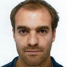
Les médecins membres du réseau TéléDiag.

Nom : FINAS
Prénom : Mathieu
Spécialités d'organe : Cardiaque, Imagerie d'Urgence, Imagerie Oncologique, Vasculaire
Numéro d'inscription à l'ordre : N° 38/11428
Fonction : Membre
Région : Auvergne-Rhône-Alpes
Etablissement(s) : CHU de Grenoble
CURSUS PROFESSIONNEL
2018-19 : Clinicat, CHU Grenoble Alpes, imagerie cardiovasculaire et digestive, diagnostique et interventionnelle.
2012 – 2017, internat radiodiagnostic et imagerie médicale en secteurs : ostéo-articulaire, thoracique, cardio-vasculaire diagnostique et interventionnel, digestive, neuroradiologie, imagerie des urgences, radiopédiatrie (CHUGA) . Stage recherche en IRM cardiaque (CHRU La Timone, Marseille) . Pneumologie et oncologie thoracique (CHUGA)
2012 : Examen Classant National : classé 974/7658
2006 – 2012 : externe des hôpitaux de Dijon, Université de Bourgogne
2006 : admis PCEM 1, classé 32/1070
2003-2004 : Année en classe préparatoire, section MPSI, Lycée Carnot, Dijon
2003 : Baccalauréat série S, spécialité mathématiques
CURSUS UNIVERSITAIRE
2018-19 : Chef de Clinique à la Faculté de Médecine (UJF)
2017 : Master 2 ISM Modèles Innovation Technologiques et Imagerie (MITI, UJF)
2016 : DIU d’imagerie cardiovasculaire diagnostique
2008-2009 : Certificat d'immunologie, Université de Bourgogne (Pr BONOTTE)
CONTRIBUTIONS
S Bricq, J Frandon, M Bernard, M Guye, M Finas, L Marcadet, L Miquerol, F Kober, G Habib, D Fagret, A Jacquier, and A Lalande, “Semiautomatic detection of myocardial contours in order to investigate normal values of the left ventricular trabeculated mass using MRI: Quantification of Trabeculated LV Mass,” J. Magn. Reson. Imaging, vol. 43, no. 6, pp. 1398–1406, Jun. 2016.
andon J, Bricq S, Bentatou Z, Marcadet L, Barral PA, Finas M, Fagret D, Kober F, Habib G, Bernard M, Lalande A, Miquerol L and Jacquier A, “Semi-automatic detection of myocardial trabeculation using cardiovascular magnetic resonance: correlation with histology and reproducibility in a mouse model of non-compaction” J Cardiovasc Magn Reson. 2018 Oct 25;20(1):70. doi: 10.1186/s12968-018-0489-0.
Bentatou Z, Finas M, Habert P, Kober F, Guye M, Bricq S, Lalande A, Frandon J, Dacher JN, Dubourg B, Habib G, Caudron J, Normant S, Rapacchi S, Bernard M and Jacquier A, “Distribution of left ventricular trabeculation across age and gender in 140 healthy Caucasian subjects on MR imaging”, Diagn Interv Imaging. 2018 Sep 24. pii: S2211-5684(18)30221-3. doi: 10.1016/j.diii.2018.08.014.
A paraître : Finas et al., “Quantification of left ventricle trabeculae in Hypertrophied Cardiomyopathy, in comparison with healthy subjects”, these de doctorat de médecine.
« Radeos » : Livret de l'interne de radiologie. Co-rédaction du chapitre « imagerie des urgences digestives », paru en octobre 2016.
COMMUNICATIONS ORALES
JFR 2016, Paris : “ Traitement endovasculaire des dissections aortiques de type B : résultats d’une cohorte mono centrique M. Finas, M. Rodière, V.Bach, N. Huet, G. Ferretti, F. Thony
JFR 2015, Paris : “Evaluation automatisée en IRM de la non compaction du ventricule gauche chez un modèle murin. Comparaison à l’histologie.” Finas M., Frandon J., Bricq ., Lalande A., Marcadet L., Miquerol L., Bernard M., Jacquier A.
ESMRMB 2015, Edinburgh : “ls it worth suppressing blood from trabeculae for the calculation of the non-compacted mass in cardiac MRI?” Finas M, Frandon J, Bricq S, Lalande A, Bernard M Jacquier A.